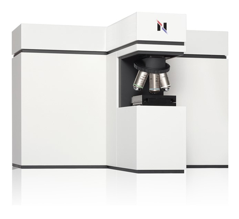
Raman asbestos fiber analysis conducted with the RAMANtouch, identifies asbestos and evaluates its potential risk. Thanks to its high spatial resolution, the RAMANtouch can detect even the smallest asbestos fibers. This level of precision ensures accurate assessment and effective management of asbestos-related risks.
Breathtaking Asbestos
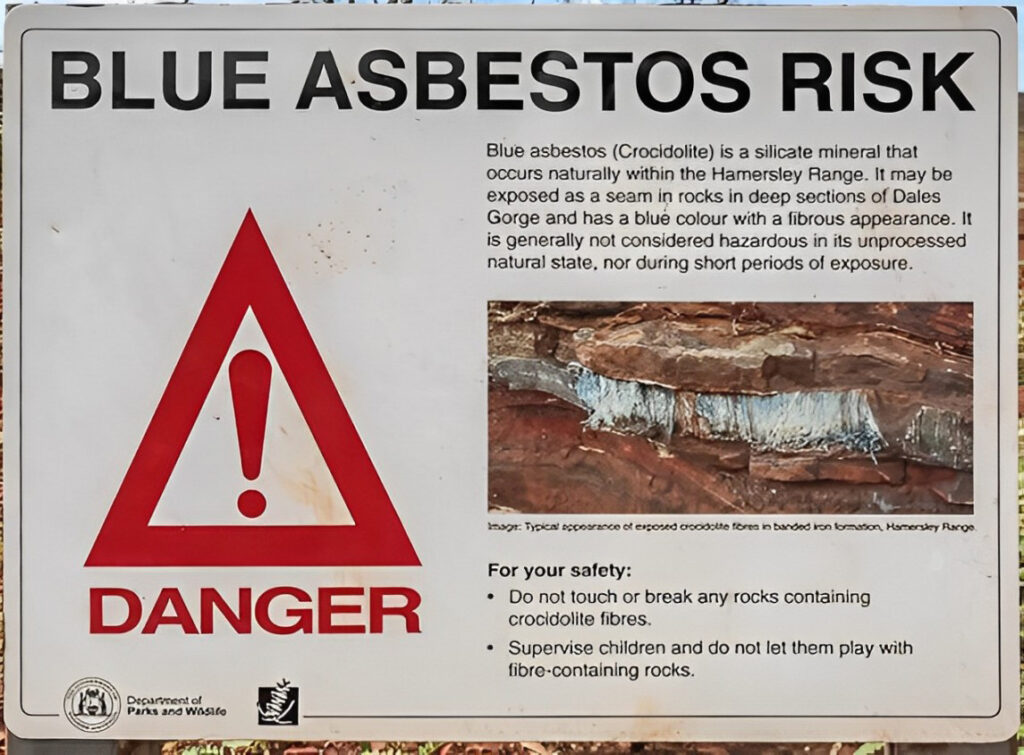
Asbestos, a naturally occurring mineral, gained widespread use throughout the 20th century due to its remarkable heat resistance and durability. Its versatility led to its incorporation into various industries, including construction, shipbuilding, and automotive manufacturing.
Asbestos challenge
Despite its utility, asbestos carries a significant health risk. When processed and handled, asbestos fibers can become airborne and easily inhaled.
Once lodged in the lungs, these microscopic fibers can cause severe health issues, including lung cancer, asbestosis, and mesothelioma. Consequently, regulations have been implemented to limit its usage and mitigate exposure.
Efforts to remove asbestos from older structures to replace it with safer alternatives are ongoing. The challenge? Asbestos fibers are sometimes hidden within other materials, and similar to non-asbestos substances.

Therefore, specialized companies are commissioned to identify asbestos using elaborate analytical techniques such as Scanning Electron Microscopy (SEM) or X-ray diffraction (XRD).
While these methods are the standard choice for asbestos identification they need extensive sample preparation and highly trained staff. Besides, SEM and XRD have further limitations.
SEM provides element maps but lacks mineral identification, while XRD identifies minerals but doesn’t offer imaging. Yet, it is crucial to both identify the respective asbestos mineral and determine its fiber’s dimensions.
A technique that is able to bridge this gap is Raman microscopy, a somewhat overlooked method in asbestos analysis. With its ability to provide both substance identification and imaging capabilities, Raman microscopy presents a viable alternative to SEM and XRD.
Raman asbestos fiber analysis
This is where Bruker’s new acquisition, the RAMANtouch, comes into play. By providing high-resolution in Raman imaging, it is able to analyze and depict the smallest of fibers down to the nanometer range. Let’s demonstrate the capabilities of the RAMANtouch with an example.
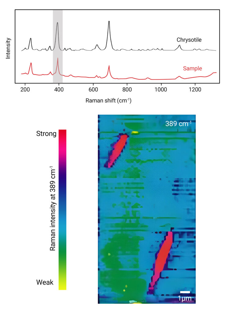
A sample was taken in a building in which fire-resistant material was used that could contain asbestos. The spectrum gathered of the sample was compared with a spectral library and revealed that the material is indeed made of chrysotile, a type of asbestos.
In addition to identifying the fiber it was also imaged, which is crucial for determining the contained fibers’ dimensions. The importance of this lies in the criteria for a material to be classified as asbestos.
The material must exhibit a fibrous shape, with a length exceeding 5 µm, a diameter of 3 µm, and a length-to-width ratio of at least 3:1.
For the fiber dimension determination, the peak at 389 cm⁻¹, indicative of chrysotile, was used to generate the color-coded map seen below with each color representing the peak’s intensity at different locations in the sample.
The picture illustrates why asbestos is so dangerous. Even though the chrysotile depicted here is considered less hazardous than the thinner and longer crocidolite, the fibrous form with sharp ends is still evident.
Conclusion
The analysis with the RAMANtouch has clearly identified the material from the building as asbestos. With knowledge of the chemical composition and dimensions of the fibers, safe disposal of the material can now take place.
Detecting and quantifying harmful mineral particles is an important job for our devices. If you want to learn more about this, have a look at this blog article about coal mining dust: